
Electromyography (EMG) is a diagnostic procedure to assess the health of muscles and the nerve cells that control them (motor neurons). Motor neurons transmit electrical signals that cause muscles to contract. An EMG translates these signals into graphs, sounds or numerical values that a specialist interprets. An EMG uses tiny devices called electrodes to transmit or detect electrical signals. During a needle EMG, a needle electrode inserted directly into a muscle records the electrical activity in that muscle. A nerve conduction study, another part of an EMG, uses electrodes taped to the skin (surface electrodes) to measure the speed and strength of signals traveling between two or more points. EMG results can reveal nerve dysfunction, muscle dysfunction or problems with nerve-to-muscle signal transmission.
Why Is an Electromyography Performed?
Your doctor may perform an EMG if you’re experiencing symptoms that may indicate a muscle or nerve disorder. Some symptoms that may call for an EMG include:
tingling
numbness
muscle weakness
muscle pain or cramping
paralysis
involuntary muscle twitching (or tics)
The results of an EMG can help your doctor determine the underlying cause of these symptoms. Possible causes could include:
muscle disorders, such as muscular dystrophy
disorders that affect the ability of the motor neuron to send electrical signals to the muscle, such as myasthenia gravis
radiculopathies
peripheral nerve disorders that affect the nerves outside the spinal cord, such as carpal tunnel syndrome
nerve disorders, such as amyotrophic lateral sclerosis (ALS)
Before the procedure:
Your doctor will explain the procedure to you and offer you the opportunity to ask any questions that you might have about the procedure.
Generally, fasting is not required before the test. In some cases, cigarettes and caffeinated beverages, such as coffee, tea, and cola may be restricted two to three hours before testing.
Notify your doctor of all medications (prescribed and over-the-counter) and herbal supplements that you are taking.
Notify your doctor if you have a pacemaker.
Dress in clothes that permit access to the area to be tested or that are easily removed.
Stop using lotions or oils on your skin for a few days before your procedure, or at least stop using them on the day of the exam.
Based on your medical condition, your doctor may request other specific preparation.
What Happens During an Electromyography?
You will be asked to lie down on an examination table or to sit in a reclined chair. Your doctor may ask you to move into different positions during the procedure.
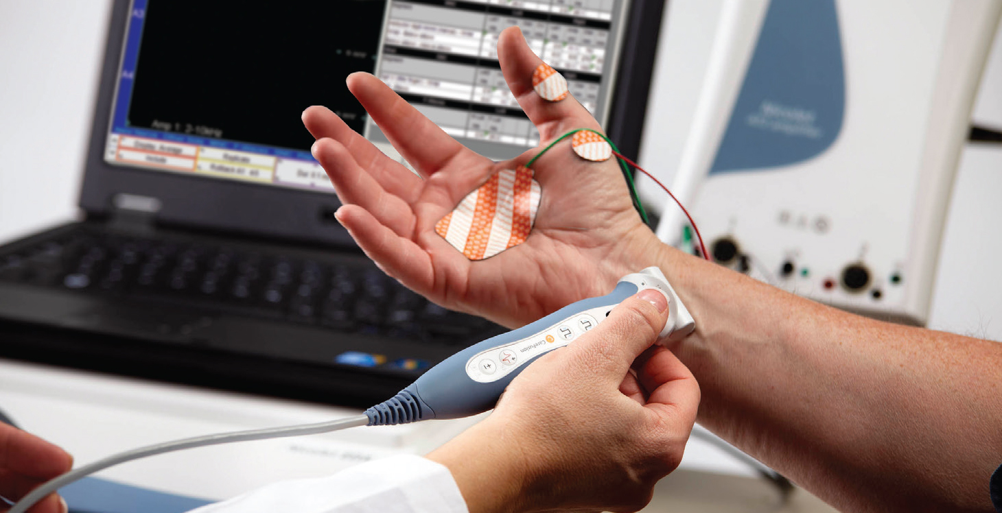
An EMG has two parts: the nerve conduction study and the needle EMG. The nerve conduction study is performed first. During this portion of the procedure, your doctor will apply several electrodes to the surface of your skin, usually in the area where you are experiencing symptoms. These electrodes will evaluate how well your motor neurons communicate with your muscles. Once the test is complete, the electrodes are removed from the skin.
After the nerve conduction study, your doctor will perform the needle EMG. Your doctor will first clean the affected area with an antiseptic. Then, they will use a needle to insert electrodes into your muscle tissue. You may feel slight discomfort or pain while the needle is being inserted.
The needle electrodes will evaluate the electrical activity of your muscles when contracted and when at rest. These electrodes will be removed after the test is over.
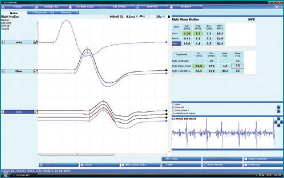
During both parts of the EMG procedure, the electrodes will deliver tiny electrical signals to your nerves. A computer will translate these signals into graphs or numerical values that can be interpreted by your doctor. The entire procedure should take between 30 and 60 minutes.
Information retrieved from http://www.mayoclinic.org, http://www.healthline.com, http://www.hopkinsmedicine.org